
מודל הוראה משולב ליולדות, מיילדות, חינוך רפואי, טיפול טרום לידתי, הכשרת מיומנויות מיילדות, סימולציה, PVC רגיל
מודל הוראה משולב ליולדות, מיילדות, חינוך רפואי, טיפול טרום לידתי, הכשרת מיומנויות מיילדות, סימולציה, PVC רגיל
F (תכונות)
מודל הכשרה זה ללידה מיילדת הוא כלי מקיף להכשרת מיומנויות מיילדות, המורכב מסימולציה של הריון
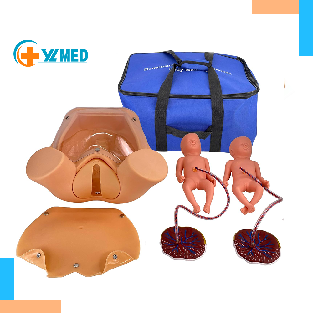
דגם פלג גוף תחתון של אישה, שני דגמי עובר, דגם שליה המחוברת לחבל הטבור ואביזרים תומכים מלאים. הוא מיועד לבדיקה טרום לידתית, מיילדות והכשרה במיומנויות לידה.
א (יתרונות)
סט אביזרים מלא מאפשר אימון לידה מלא; עיצוב עובר כפול תומך בתרגול לידה כפולה;
מבנה אגן ועובר מדויקים מבחינה אנטומית מספקים משוב מישושי אותנטי; חומר עמיד עומד בפני פעולות חוזרות ונשנות
הכשרה בניתוח; מתאים להכשרה טכנית מיילדותית בסיסית ומתקדמת.
ב' (הטבות)
הוא משמש ככלי הכשרה חיוני למיילדות, מיילדות וסטודנטים לסיעוד כדי לשלוט בלידה, מיילדות ו...
מיומנויות בדיקה טרום לידתית, שיפור המיומנות התפעולית ובטיחות המטופל בלידה קלינית. אידיאלי עבור בדיקות רפואיות
בתי ספר, מרכזי הכשרה למיילדות ובתי חולים למיילדות וגינקולוגיה.
C (הסמכות והוכחות)
מודל זה עומד בתקני חינוך בינלאומיים למיילדות וגינקולוגיה, הנמצאים בשימוש נרחב במוסדות הכשרה למיילדות ברחבי העולם, ומוכרים בזכות הסימולציה הריאליסטית והערך החינוכי המעשי שלו.
מודל הכשרה זה ללידה מיילדת הוא כלי מקיף להכשרת מיומנויות מיילדות, המורכב מסימולציה של הריון
דגם פלג גוף תחתון של אישה, שני דגמי עובר, דגם שליה המחוברת לחבל הטבור ואביזרים תומכים מלאים. הוא מיועד לבדיקה טרום לידתית, מיילדות והכשרה במיומנויות לידה.
א (יתרונות)
סט אביזרים מלא מאפשר אימון לידה מלא; עיצוב עובר כפול תומך בתרגול לידה כפולה;
מבנה אגן ועובר מדויקים מבחינה אנטומית מספקים משוב מישושי אותנטי; חומר עמיד עומד בפני פעולות חוזרות ונשנות
הכשרה בניתוח; מתאים להכשרה טכנית מיילדותית בסיסית ומתקדמת.
ב' (הטבות)
הוא משמש ככלי הכשרה חיוני למיילדות, מיילדות וסטודנטים לסיעוד כדי לשלוט בלידה, מיילדות ו...
מיומנויות בדיקה טרום לידתית, שיפור המיומנות התפעולית ובטיחות המטופל בלידה קלינית. אידיאלי עבור בדיקות רפואיות
בתי ספר, מרכזי הכשרה למיילדות ובתי חולים למיילדות וגינקולוגיה.
C (הסמכות והוכחות)
מודל זה עומד בתקני חינוך בינלאומיים למיילדות וגינקולוגיה, הנמצאים בשימוש נרחב במוסדות הכשרה למיילדות ברחבי העולם, ומוכרים בזכות הסימולציה הריאליסטית והערך החינוכי המעשי שלו.
מודל ליולדות לנשים בהריון להוראה מיילדותית לבדיקה טרום לידתית, מודל מיומנויות ליולדות בהכשרת מיילדות
תיאור מוצר

פרטים
מודל מקיף להכשרת מיומנויות למסירה
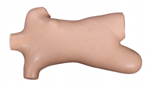
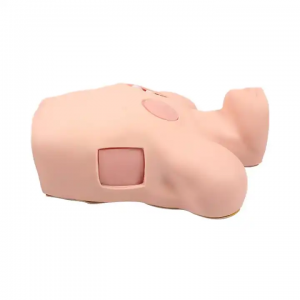
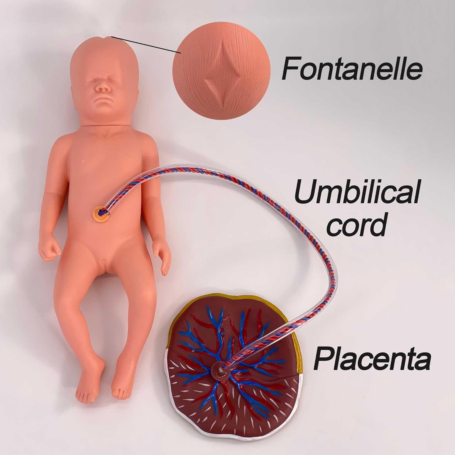
המודל הורכב ממודל פלג גוף תחתון מדומה של נשים בהריון, מודל של שני עוברים, מודל המחובר לחבל טבור ומודל של שליה.
מוצר זה מיועד להכשרה טכנית בסיסית במיילדות, ומספק תרגילים מקיפים במיומנויות כגון בדיקה טרום לידתית, מיילדות ולידה.
תכונת מוצר

תכונות פונקציונליות:
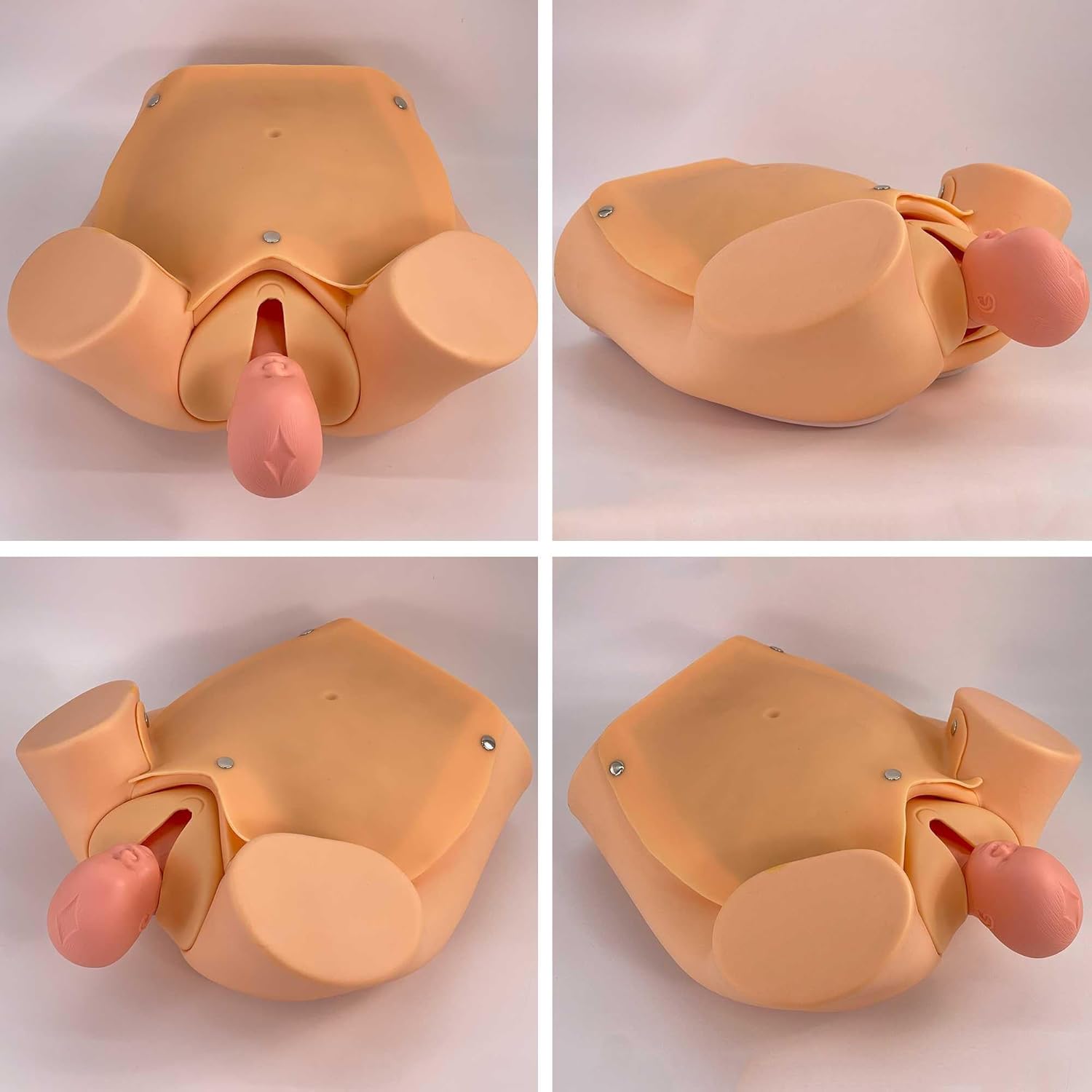
■ מודל לבדיקת צוואר הרחם: נקבעו גודל התרחבות פתח צוואר הרחם, מידת השינוי בפתח צוואר הרחם ויחסי המיקום בין ראש העובר למישור עמוד השדרה השת. שלב 1: פתח צוואר הרחם לא הורחב, תעלת צוואר הרחם לא אבדה, ויחסי המיקום בין ראש העובר למישור עמוד השדרה השת היו -5. שלב 2: פתח צוואר הרחם הורחב ב-2 ס"מ, תעלת צוואר הרחם נעלמה ב-50%, ויחסי המיקום בין ראש העובר למישור עמוד השדרה השת היו -4. שלב 3: פתח צוואר הרחם הורחב ב-4 ס"מ, תעלת צוואר הרחם נעלמה לחלוטין, ויחסי המיקום בין ראש העובר למישור עמוד השדרה השת היו -3. שלב 4: פתח צוואר הרחם הורחב ב-5 ס"מ, תעלת צוואר הרחם נעלמה לחלוטין, ויחס המיקום בין ראש העובר למישור עמוד השדרה השת היה 0. שלב 5: פתח צוואר הרחם הורחב ב-7 ס"מ, תעלת צוואר הרחם נעלמה לחלוטין, ויחס המיקום בין ראש העובר למישור עמוד השדרה השת היה +2. שלב 6: פתח צוואר הרחם הורחב ב-10 ס"מ, תעלת צוואר הרחם נעלמה לחלוטין, ויחס המיקום בין ראש העובר למישור עמוד השדרה השת היה +5. ■ מודל עובר: עורו של עובר סטנדרטי רך, ניתן להבחין בפונטנלים, וניתן לתרגל שאיבה ראשונית. אביזרים עובריים: חבל טבור, שליה. ■ מודל לידה: חיץ הנקבים של המודל רך וגמיש, ויכול לדמות את פעולת המיילדות במצב אמיתי. ניתן לתרגל טכניקת הנקה של חיץ הנקבים. הבטן הורכבה מדופן בטן שקופה ועור מדומה, דבר שהיה נוח לצפייה בהדגמת תפקוד הלידה וביחסי המיקום המישוריים בין ראש העובר לעמוד השדרה הסיאטי. ■ מודל תפר פרינאום: פתחי האפיזיוטומיה של המודל היו כדלקמן:
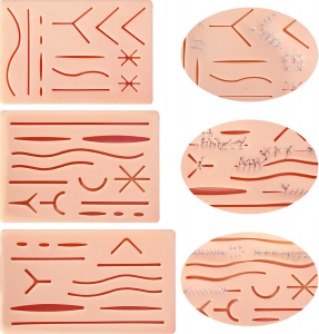
חתך אמצעי, חתך שמאלי וחתך ימני. ניתן לבצע שלושה סוגים של תרגילי תפירה לחתך פרינאום.